Niosome
A niosome is a non-ionic surfactant-based vesicle. Niosomes are formed mostly by non-ionic surfactant and cholesterol incorporation as an excipient.[1] Other excipients can also be used. Niosomes have more penetrating capability than the previous preparations of emulsions.[2] They are structurally similar to liposomes in having a bilayer, however, the materials used to prepare niosomes make them more stable. It can entrap both hydrophilic and lipophilic drugs, either in an aqueous layer or in a vesicular membrane made of lipid material.

Structure
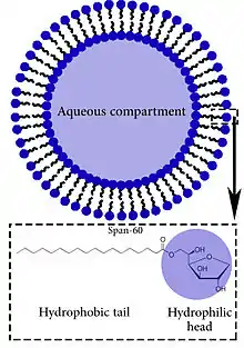
Niosomes are lamellar structures that are microscopic in size. They constitute of non-ionic surfactant of the alkyl or dialkyl polyglycerol ether class and cholesterol with subsequent hydration in aqueous media. The surfactant molecules tend to orient themselves in such a way that the hydrophilic ends of the non-ionic surfactant point outwards, while the hydrophobic ends face each other to form the bilayer. The figure in this article on niosomes gives a better idea of the lamellar orientation of the surfactant molecules.[3]
Advantages of niosomes
- Niosomes are osmotically active, chemically stable and have long storage time compared to liposomes
- Their surface formation and modification is very easy because of the functional groups on their hydrophilic heads
- They have high compatibility with biological systems and low toxicity because of their non-ionic nature
- They are biodegradable and non-immunogenic
- They can entrap lipophilic drugs into vesicular bilayer membranes and hydrophilic drugs in aqueous compartments
- They can improve the therapeutic performance of the drug molecules by protecting the drug from biological environment, resulting in better availability and controlled drug delivery by restricting the drug effects to target cells in targeted carriers and delaying clearance from the circulation in sustained drug delivery
- Access to raw materials is convenient [1]
Methods of preparation
Niosomes can be prepared by various methods, including:[1]
- Ether injection method (EIM)
- Hand shaking method (HSM)
- Reverse phase evaporation method (REV)
- trans membrane pH gradient
- the "Bubble" method
- Microfluidization method
- formation of niosomes from proniosomes (Proniosome technology (PT))[4]
- Thin-film hydration method (TFH)
- Heating method (HM)
- Freeze and thaw method (FAT)
- Dehydration rehydration method (DRM)
Applications
Niosomes are a novel drug delivery system that are finding application in:
References
- Moghassemi, Saeid; Hadjizadeh, Afra (2014). "Nano-niosomes as nanoscale drug delivery systems: An illustrated review". Journal of Controlled Release. 185: 22–36. doi:10.1016/j.jconrel.2014.04.015. PMID 24747765.
- Paracha, Usman Zafar (14 May 2008). "Your Source of Information: Niosomes".
- PharmaXChange.info - Niosomes Article on niosomes covering topics such as structure, methods of preparation, advantages and applications
- "PharmaXChange.info - Articles - Niosomes".
- Moghassemi, Saeid, and Afra Hadjizadeh. "Nano-niosomes as nanoscale drug delivery systems: an illustrated review." Journal of Controlled Release 185 (2014): 22-36.
- Puras, G., M. Mashal, J. Zárate, M. Agirre, E. Ojeda, S. Grijalvo, R. Eritja et al. "A novel cationic niosome formulation for gene delivery to the retina." Journal of Controlled Release 174 (2014): 27-36.
- "Niosomes".
- "Cosmetic and pharmaceutical compositions containing niosomes and a water-soluble polyamide, and a process for preparing these compositions".