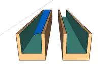
Open microfluidics
Microfluidics refers to the flow of fluid in channels or networks with at least one dimension on the micron scale.[1][2] In open microfluidics, also referred to as open surface microfluidics or open-space microfluidics, at least one boundary confining the fluid flow of a system is removed, exposing the fluid to air or another interface such as a second fluid.[1][3][4]
Types of open microfluidics
Open microfluidics can be categorized into various subsets. Some examples of these subsets include open-channel microfluidics, paper-based, and thread-based microfluidics.[1][5][6]
Open-channel microfluidics
In open-channel microfluidics, a surface tension-driven capillary flow occurs and is referred to as spontaneous capillary flow (SCF).[1][7] SCF occurs when the pressure at the advancing meniscus is negative.[1] The geometry of the channel and contact angle of fluids has been shown to produce SCF if the following equation is true.
Where pf is the free perimeter of the channel (i.e., the interface not in contact with the channel wall), and pw is the wetted perimeter[8] (i.e., the walls in contact with the fluid), and θ is the contact angle of the fluid on the material of the device.[1][5]
Paper-based microfluidics
Paper-based microfluidics utilizes the wicking ability of paper for functional readouts.[9][10] Paper-based microfluidics is an attractive method because paper is cheap, easily accessible, and has a low environmental impact. Paper is also versatile because it is available in various thicknesses and pore sizes.[9] Coatings such as wax have been used to guide flow in paper microfluidics.[11] In some cases, dissolvable barriers have been used to create boundaries on the paper and control the fluid flow.[12] The application of paper as a diagnostic tool has shown to be powerful because it has successfully been used to detect glucose levels,[13] bacteria,[14] viruses,[15] and other components in whole blood.[16] Cell culture methods within paper have also been developed.[17][18] Lateral flow immunoassays, such as those used in pregnancy tests, are one example of the application of paper for point of care or home-based diagnostics.[19] Disadvantages include difficulty of fluid retention and high limits of detection.
Thread-based microfluidics
Thread-based microfluidics, an offshoot from paper-based microfluidics, utilizes the same capillary based wicking capabilities.[20] Common thread materials include nitrocellulose, rayon, nylon, hemp, wool, polyester, and silk.[21] Threads are versatile because they can be woven to form specific patterns.[22] Additionally, two or more threads can converge together in a knot bringing two separate ‘streams’ of fluid together as a reagent mixing method.[23] Threads are also relatively strong and difficult to break from handling which makes them stable over time and easy to transport.[21] Thread-based microfluidics has been applied to 3D tissue engineering and analyte analysis.[24][20]
Capillary filaments in open microfluidics
Open capillary microfluidics are channels that expose fluids to open air by excluding the ceiling and/or floor of the channel.[5] Rather than rely on using pumps or syringes to maintain flow, open capillary microfluidics uses surface tension to facilitate the flow.[25] The elimination of and infusion source reduces the size of the device and associated apparatus, along with other aspects that could obstruct their use. The dynamics of capillary-driven flow in open microfluidics are highly reliant on two types of geometric channels commonly known as either rectangular U-grooves or triangular V-grooves.[26][25] The geometry of the channels dictates the flow along the interior walls fabricated with various ever-evolving processes.[27]
Capillary filaments in U-groove

Rectangular open-surface U-grooves are the easiest type of open microfluidic channel to fabricate. This design can maintain the same order of magnitude velocity in comparison to V-groove.[28][26][29] Channels are made of glass or high clarity glass substitutes such as polymethyl methacrylate (PMMA), polycarbonate (PC), or cyclic olefin copolymer (COP). To eliminate the remaining resistance after etching, channels are given hydrophilic treatment using oxygen plasma or deep reactive-ion etching(DRIE).[30][31][32]
Capillary filaments in V-groove

V-groove, unlike U-groove, allows for a variety of velocities depending on the groove angle.[29] V-grooves with sharp groove angle result in the interface curvature at the corners explained by reduced Concus-Finn conditions.[33] In a perfect inner corner of a V-groove, the filament will advance indefinitely in the groove allowing the formation of capillary filament depending on the wetting conditions.[34] The width of the groove plays an important role in controlling the fluid flow. The narrower the V-groove is, the better the capillary flow of liquids is even for highly viscous liquids such as blood; this effect has been used to produce an autonomous assay.[5][35] The fabrication of a V-groove is more difficult than a U-groove as it poses a higher risk for faulty construction, since the corner has to be tightly sealed.[30]
Advantages
One of the main advantages of open microfluidics is ease of accessibility which enables intervention (i.e., for adding or removing reagents) to the flowing liquid in the system.[36] Open microfluidics also allows simplicity of fabrication thus eliminating the need to bond surfaces. When one of the boundaries of a system is removed, a larger liquid-gas interface results, which enables liquid-gas reactions.[1][37] Open microfluidic devices enable better optical transparency because at least one side of the system is not covered by the material which can reduce autofluorescence during imaging.[38] Further, open systems minimize and sometimes eliminate bubble formation, a common problem in closed systems.[1]
In closed system microfluidics, the flow in the channels is driven by pressure via pumps (syringe pumps), valves (trigger valves), or electrical field.[39] An example of one of these methods for achieving low flow rates using temperature-controlled evaporation has been described for an open microfluidics system, allowing for long incubation hours for biological applications and requiring small sample volumes.[40] Open system microfluidics enable surface-tension driven flow in channels thereby eliminating the need for external pumping methods.[36][41] For example, some open microfluidic devices consist of a reservoir port and pumping port that can be filled with fluid using a pipette.[1][5][36] Eliminating external pumping requirements lowers cost and enables device use in all laboratories with pipettes.[37]
Disadvantages
Some drawbacks of open microfluidics include evaporation,[42] contamination,[43] and limited flow rate.[4] Open systems are susceptible to evaporation which can greatly affect readouts when fluid volumes are on the microscale.[42] Additionally, due to the nature of open systems, they are more susceptible to contamination than closed systems.[43] Cell culture and other methods where contamination or small particulates are a concern must be carefully performed to prevent contamination. Lastly, open systems have a limited flow rate because induced pressures cannot be used to drive flow.[4]
Applications
Like many microfluidic technologies, open system microfluidics has been applied to nanotechnology, biotechnology, fuel cells, and point of care (POC) testing.[1][4][44] For cell-based studies, open-channel microfluidic devices enable access to cells for single cell probing within the channel.[45] Other applications include capillary gel electrophoresis, water-in-oil emulsions, and biosensors for POC systems.[3][46][47] Suspended microfluidic devices, open microfluidic devices where the floor of the device is removed, have been used to study cellular diffusion and migration of cancer cells.[5] Suspended and rail-based microfluidics have been used for micropatterning and studying cell communication.[1]
References
- Berthier J (2016). Open microfluidics. Brakke, Kenneth A., Berthier, Erwin. Hoboken, NJ: Wiley. ISBN 9781118720936. OCLC 953661963.
- Whitesides GM (July 2006). "The origins and the future of microfluidics". Nature. 442 (7101): 368–73. Bibcode:2006Natur.442..368W. doi:10.1038/nature05058. PMID 16871203. S2CID 205210989.
- Pfohl T, Mugele F, Seemann R, Herminghaus S (December 2003). "Trends in microfluidics with complex fluids". ChemPhysChem. 4 (12): 1291–8. doi:10.1002/cphc.200300847. PMID 14714376.
- Kaigala GV, Lovchik RD, Delamarche E (November 2012). "Microfluidics in the "open space" for performing localized chemistry on biological interfaces". Angewandte Chemie. 51 (45): 11224–40. doi:10.1002/anie.201201798. PMID 23111955.
- Casavant BP, Berthier E, Theberge AB, Berthier J, Montanez-Sauri SI, Bischel LL, et al. (June 2013). "Suspended microfluidics". Proceedings of the National Academy of Sciences of the United States of America. 110 (25): 10111–6. Bibcode:2013PNAS..11010111C. doi:10.1073/pnas.1302566110. PMC 3690848. PMID 23729815.
- Yamada K, Shibata H, Suzuki K, Citterio D (March 2017). "Toward practical application of paper-based microfluidics for medical diagnostics: state-of-the-art and challenges". Lab on a Chip. 17 (7): 1206–1249. doi:10.1039/c6lc01577h. PMID 28251200.
- Yang D, Krasowska M, Priest C, Popescu MN, Ralston J (2011-09-07). "Dynamics of Capillary-Driven Flow in Open Microchannels". The Journal of Physical Chemistry C. 115 (38): 18761–18769. doi:10.1021/jp2065826. ISSN 1932-7447.
- "Wetted perimeter", Wikipedia, 2018-11-27, retrieved 2019-04-16
- Hosseini S, Vázquez-Villegas P, Martínez-Chapa SO (2017-08-22). "Paper and Fiber-Based Bio-Diagnostic Platforms: Current Challenges and Future Needs". Applied Sciences. 7 (8): 863. doi:10.3390/app7080863.
- Swanson C, Lee S, Aranyosi A, Tien B, Chan C, Wong M, Lowe J, Jain S, Ghaffari R (2015-09-01). "Rapid light transmittance measurements in paper-based microfluidic devices". Sensing and Bio-Sensing Research. 5: 55–61. doi:10.1016/j.sbsr.2015.07.005. ISSN 2214-1804.
- Müller RH, Clegg DL (September 1949). "Automatic Paper Chromatography". Analytical Chemistry. 21 (9): 1123–1125. doi:10.1021/ac60033a032. ISSN 0003-2700.
- Fu E, Lutz B, Kauffman P, Yager P (April 2010). "Controlled reagent transport in disposable 2D paper networks". Lab on a Chip. 10 (7): 918–20. doi:10.1039/b919614e. PMC 3228840. PMID 20300678.
- Martinez AW, Phillips ST, Carrilho E, Thomas SW, Sindi H, Whitesides GM (May 2008). "Simple telemedicine for developing regions: camera phones and paper-based microfluidic devices for real-time, off-site diagnosis". Analytical Chemistry. 80 (10): 3699–707. doi:10.1021/ac800112r. PMC 3761971. PMID 18407617.
- Shih CM, Chang CL, Hsu MY, Lin JY, Kuan CM, Wang HK, et al. (December 2015). "Paper-based ELISA to rapidly detect Escherichia coli". Talanta. 145: 2–5. doi:10.1016/j.talanta.2015.07.051. PMID 26459436.
- Wang H, Tsai C, Chen K, Tang C, Leou J, Li P, Tang Y, Hsieh H, Wu H (February 2014). "Immunoassays: Cellulose-Based Diagnostic Devices for Diagnosing Serotype-2 Dengue Fever in Human Serum (Adv. Healthcare Mater. 2/2014)". Advanced Healthcare Materials. 3 (2): 154. doi:10.1002/adhm.201470008. ISSN 2192-2640.
- Yang X, Forouzan O, Brown TP, Shevkoplyas SS (January 2012). "Integrated separation of blood plasma from whole blood for microfluidic paper-based analytical devices". Lab on a Chip. 12 (2): 274–80. doi:10.1039/c1lc20803a. PMID 22094609.
- Tao FF, Xiao X, Lei KF, Lee I (2015-03-18). "Paper-based cell culture microfluidic system". BioChip Journal. 9 (2): 97–104. doi:10.1007/s13206-015-9202-7. ISSN 1976-0280. S2CID 54718125.
- Walsh DI, Lalli ML, Kassas JM, Asthagiri AR, Murthy SK (June 2015). "Cell chemotaxis on paper for diagnostics". Analytical Chemistry. 87 (11): 5505–10. doi:10.1021/acs.analchem.5b00726. PMID 25938457.
- Lam T, Devadhasan JP, Howse R, Kim J (April 2017). "A Chemically Patterned Microfluidic Paper-based Analytical Device (C-µPAD) for Point-of-Care Diagnostics". Scientific Reports. 7 (1): 1188. Bibcode:2017NatSR...7.1188L. doi:10.1038/s41598-017-01343-w. PMC 5430703. PMID 28446756.
- Erenas MM, de Orbe-Payá I, Capitan-Vallvey LF (May 2016). "Surface Modified Thread-Based Microfluidic Analytical Device for Selective Potassium Analysis". Analytical Chemistry. 88 (10): 5331–7. doi:10.1021/acs.analchem.6b00633. PMID 27077212.
- Reches M, Mirica KA, Dasgupta R, Dickey MD, Butte MJ, Whitesides GM (June 2010). "Thread as a matrix for biomedical assays". ACS Applied Materials & Interfaces. 2 (6): 1722–8. CiteSeerX 10.1.1.646.8048. doi:10.1021/am1002266. PMID 20496913.
- Li X, Tian J, Shen W (January 2010). "Thread as a versatile material for low-cost microfluidic diagnostics". ACS Applied Materials & Interfaces. 2 (1): 1–6. doi:10.1021/am9006148. PMID 20356211.
- Ballerini DR, Li X, Shen W (March 2011). "Flow control concepts for thread-based microfluidic devices". Biomicrofluidics. 5 (1): 14105. doi:10.1063/1.3567094. PMC 3073008. PMID 21483659.
- Mostafalu P, Akbari M, Alberti KA, Xu Q, Khademhosseini A, Sonkusale SR (2016-07-18). "A toolkit of thread-based microfluidics, sensors, and electronics for 3D tissue embedding for medical diagnostics". Microsystems & Nanoengineering. 2 (1): 16039. doi:10.1038/micronano.2016.39. PMC 6444711. PMID 31057832.
- Berthier J, Brakke KA, Gosselin D, Navarro F, Belgacem N, Chaussy D (July 2016). "Spontaneous capillary flow in curved, open microchannels". Microfluidics and Nanofluidics. 20 (7): 100. doi:10.1007/s10404-016-1766-6. ISSN 1613-4982.
- Berthier J, Brakke KA, Gosselin D, Huet M, Berthier E (2014). "Metastable capillary filaments in rectangular cross-section open microchannels". AIMS Biophysics. 1 (1): 31–48. doi:10.3934/biophy.2014.1.31. ISSN 2377-9098.
- Yang D, Krasowska M, Priest C, Popescu MN, Ralston J (2011-09-29). "Dynamics of Capillary-Driven Flow in Open Microchannels". The Journal of Physical Chemistry C. 115 (38): 18761–18769. doi:10.1021/jp2065826. ISSN 1932-7447.
- Berthier J, Brakke KA, Gosselin D, Bourdat AG, Nonglaton G, Villard N, et al. (2014-09-18). "Suspended microflows between vertical parallel walls". Microfluidics and Nanofluidics. 18 (5–6): 919–929. doi:10.1007/s10404-014-1482-z. ISSN 1613-4982.
- Han A, Mondin G, Hegelbach NG, de Rooij NF, Staufer U (January 2006). "Filling kinetics of liquids in nanochannels as narrow as 27 nm by capillary force" (PDF). Journal of Colloid and Interface Science. 293 (1): 151–7. Bibcode:2006JCIS..293..151H. doi:10.1016/j.jcis.2005.06.037. PMID 16023663.
- Kitron-Belinkov M, Marmur A, Trabold T, Dadheech GV (July 2007). "Groovy drops: effect of groove curvature on spontaneous capillary flow". Langmuir. 23 (16): 8406–10. doi:10.1021/la700473m. PMID 17608505.
- Gambino J (2011). "Process Challenges for Integration of Copper Interconnects with Low-k Dielectrics". ECS Transactions. 35 (4). Montreal, QC, Canada: 687–699. Bibcode:2011ECSTr..35d.687G. doi:10.1149/1.3572313. Cite journal requires
|journal=(help) - Schilp A, Hausner M, Puech M, Launay N, Karagoezoglu H, Laermer F (2001). Advanced Etch Tool for High Etch Rate Deep Reactive Ion Etching in Silicon Micromachining Production Environment. Advanced Microsystems for Automotive Applications 2001. Berlin Heidelberg: Springer. pp. 229–236. ISBN 978-3-642-62124-6.
- Berthier J, Brakke KA, Berthier E (2013-11-06). "A general condition for spontaneous capillary flow in uniform cross-section microchannels". Microfluidics and Nanofluidics. 16 (4): 779–785. doi:10.1007/s10404-013-1270-1. ISSN 1613-4982.
- Yost FG, Rye RR, Mann Jr JA (December 1997). "Solder wetting kinetics in narrow V-grooves". Acta Materialia. 45 (12): 5337–5345. doi:10.1016/s1359-6454(97)00205-x. ISSN 1359-6454.
- Faivre M, Peltié P, Planat-Chrétien A, Cosnier ML, Cubizolles M, Nougier C, et al. (May 2011). "Coagulation dynamics of a blood sample by multiple scattering analysis". Journal of Biomedical Optics. 16 (5): 057001–057001–9. Bibcode:2011JBO....16e7001F. doi:10.1117/1.3573813. PMID 21639579.
- Lee JJ, Berthier J, Brakke KA, Dostie AM, Theberge AB, Berthier E (May 2018). "Droplet Behavior in Open Biphasic Microfluidics". Langmuir. 34 (18): 5358–5366. doi:10.1021/acs.langmuir.8b00380. PMID 29692173.
- Zhao B, Moore JS, Beebe DJ (February 2001). "Surface-directed liquid flow inside microchannels". Science. 291 (5506): 1023–6. Bibcode:2001Sci...291.1023Z. doi:10.1126/science.291.5506.1023. PMID 11161212.
- Young EW, Berthier E, Beebe DJ (January 2013). "Assessment of enhanced autofluorescence and impact on cell microscopy for microfabricated thermoplastic devices". Analytical Chemistry. 85 (1): 44–9. doi:10.1021/ac3034773. PMC 4017339. PMID 23249264.
- Sackmann EK, Fulton AL, Beebe DJ (March 2014). "The present and future role of microfluidics in biomedical research". Nature. 507 (7491): 181–9. Bibcode:2014Natur.507..181S. doi:10.1038/nature13118. PMID 24622198. S2CID 4459357.
- Zimmermann M, Bentley S, Schmid H, Hunziker P, Delamarche E (December 2005). "Continuous flow in open microfluidics using controlled evaporation". Lab on a Chip. 5 (12): 1355–9. doi:10.1039/B510044E. PMID 16286965.
- Brakke, Kenneth A. (2015-01-31). The Motion of a Surface by Its Mean Curvature. (MN-20). Princeton: Princeton University Press. doi:10.1515/9781400867431. ISBN 9781400867431.
- Kachel S, Zhou Y, Scharfer P, Vrančić C, Petrich W, Schabel W (February 2014). "Evaporation from open microchannel grooves". Lab on a Chip. 14 (4): 771–8. doi:10.1039/c3lc50892g. PMID 24345870.
- Ogawa M, Higashi K, Miki N (August 2015). "Development of hydrogel microtubes for microbe culture in open environment". Micromachines. 2015 (6): 5896–9. doi:10.3390/mi8060176. PMC 6190135. PMID 26737633.
- Dak P, Ebrahimi A, Swaminathan V, Duarte-Guevara C, Bashir R, Alam MA (April 2016). "Droplet-based Biosensing for Lab-on-a-Chip, Open Microfluidics Platforms". Biosensors. 6 (2): 14. doi:10.3390/bios6020014. PMC 4931474. PMID 27089377.
- Hsu CH, Chen C, Folch A (October 2004). ""Microcanals" for micropipette access to single cells in microfluidic environments". Lab on a Chip. 4 (5): 420–4. doi:10.1039/b404956j. PMID 15472724.
- Li C, Boban M, Tuteja A (April 2017). "Open-channel, water-in-oil emulsification in paper-based microfluidic devices". Lab on a Chip. 17 (8): 1436–1441. doi:10.1039/c7lc00114b. PMID 28322402.
- Gutzweiler L, Gleichmann T, Tanguy L, Koltay P, Zengerle R, Riegger L (July 2017). "Open microfluidic gel electrophoresis: Rapid and low cost separation and analysis of DNA at the nanoliter scale". Electrophoresis. 38 (13–14): 1764–1770. doi:10.1002/elps.201700001. PMID 28426159.