Oxylipin
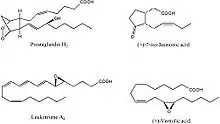
Oxylipins constitute a family of oxygenated natural products which are formed from fatty acids by pathways involving at least one step of dioxygen-dependent oxidation.[1] Oxylipins are derived from polyunsaturated fatty acids (PUFAs) by COX enzymes (cyclooxygenases), by LOX enzymes (lipoxygenases), or by cytochrome P450 epoxygenase.[2]

Oxylipins are widespread in aerobic organisms including plants, animals and fungi. Many of oxylipins have physiological significance.[3][4] Typically, oxylipins are not stored in tissues but are formed on demand by liberation of precursor fatty acids from esterified forms.
Biosynthesis
Biosynthesis of oxylipins is initiated by dioxygenases or monooxygenases; however also non-enzymatic autoxidative processes contribute to oxylipin formation (phytoprostanes, isoprostanes). Dioxygenases include lipoxygenases (plants, animals, fungi), heme-dependent fatty acid oxygenases (plants, fungi), and cyclooxygenases (animals). Fatty acid hydroperoxides or endoperoxides are formed by action of these enzymes. Monooxygenases involved in oxylipin biosynthesis are members of the cytochrome P450 superfamily and can oxidize double bonds with epoxide formation or saturated carbons forming alcohols. Nature has evolved numerous enzymes which metabolize oxylipins into secondary products, many of which possess strong biological activity. Of special importance are the cytochrome P450 enzymes in animals, including CYP5A1 (thromboxane synthase), CYP8A1 (prostacyclin synthase), and the CYP74 family of hydroperoxide-metabolizing enzymes in plants, lower animals and bacteria. In the plant and animal kingdoms the C18 and C20 polyenoic fatty acids, respectively, are the major precursors of oxylipins.
Structure and function
Oxylipins in animals, referred to as eicosanoids (Greek icosa; twenty) because of their formation from twenty-carbon essential fatty acids, have potent and often opposing effects on e.g. smooth muscle (vasculature, myometrium) and blood platelets. Certain eicosanoids (leukotrienes B4 and C4) are proinflammatory whereas others (resolvins, protectins) are antiinflammatory and are involved in the resolution process which follows tissue injury. Plant oxylipins are mainly involved in control of ontogenesis, reproductive processes and in the resistance to various microbial pathogens and other pests.
Oxylipins most often act in an autocrine or paracrine manner, notably in targeting peroxisome proliferator-activated receptors (PPARs) to modify adipocyte formation and function.[2]
Most oxylipins in the body are derived from linoleic acid or alpha-linolenic acid. Linoleic acid oxylipins are usually present in blood and tissue in higher concentrations than any other PUFA oxylipin, despite the fact that alpha-linolenic acid is more readily metabolized to oxylipin.[5]
Linoleic acid oxylipins can be anti-inflammatory, but are more often pro-inflammatory, associated with atherosclerosis, non-alcoholic fatty liver disease, and Alzheimer's disease.[5] Centenarians have shown reduced levels of linoleic acid oxylipins in their blood circulation.[6] Lowering dietary linoleic acid results in fewer linoleic acid oxylipins in humans.[7] From 1955 to 2005 the linoleic acid content of human adipose tissue has risen an estimated 136% in the United States.[8]
In general, oxylipins derived from omega-6 fatty acids are more pro-inflammatory, vasconstrictive, and proliferative than those derived from omega-3 fatty acids.[5] The omega-3 eicosapentaenoic acid (EPA)-derived and docosahexaenoic acid (DHA)-derived oxylipins are anti-inflammatory and vasodilatory.[5] In a clinical trial of men with high triglycerides, 3 grams daily of DHA compared with placebo (olive oil) given for 91 days nearly tripled the DHA in red blood cells while reducing oxylipins in those cells.[9] Both groups were given Vitamin C (ascorbyl palmitate) and Vitamin E (mixed tocopherol) supplements.[9]
References
- Wasternack C (2007). "Jasmonates: an update on biosynthesis, signal transduction and action in plant stress response, growth and development". Annals of Botany. 100 (4): 681–697. doi:10.1093/aob/mcm079. PMC 2749622. PMID 17513307.
- Barquissau V, Ghandour RA, Ailhaud G, Klingenspor M, Langin D, Amri EZ, Pisani DF (2017). "Control of adipogenesis by oxylipins, GPCRs and PPARs". Biochimie. 136: 3–11. doi:10.1016/j.biochi.2016.12.012. PMID 28034718.
- Zhao J, Davis LC, Verpoorte R (2005). "Elicitor signal transduction leading to production of plant secondary metabolites". Biotechnol. Adv. 23 (4): 283–333. doi:10.1016/j.biotechadv.2005.01.003. PMID 15848039.
- Bolwell GP, Bindschedler LV, Blee KA, Butt VS, Davies DR, Gardner SL, Gerrish C, Minibayeva F (2002). "The apoplastic oxidative burst in response to biotic stress in plants: a three-component system". J. Exp. Bot. 53 (372): 1367–1376. doi:10.1093/jexbot/53.372.1367. PMID 11997382.
- Gabbs M, Leng S, Devassy JG, Monirujjaman M, Aukema HM (2015). "Advances in Our Understanding of Oxylipins Derived from Dietary PUFAs". Advances in Nutrition. 6 (5): 513–540. doi:10.3945/an.114.007732. PMC 4561827. PMID 26374175.
- Collino S, Montoliu I, Martin FP, Scherer M, Mari D, Salvioli S, Bucci L, Ostan R, Monti D, Biagi E, Brigidi P, Franceschi C, Rezzi S (2013). "Metabolic signatures of extreme longevity in northern Italian centenarians reveal a complex remodeling of lipids, amino acids, and gut microbiota metabolism". PLOS ONE. 8 (3): e56564. Bibcode:2013PLoSO...856564C. doi:10.1371/journal.pone.0056564. PMC 3590212. PMID 23483888.
- Ramsden CE, Ringel A, Feldstein AE, Taha AY, MacIntosh BA, Hibbeln JR, Majchrzak-Hong SF, Faurot KR, Rapoport SI, Cheon Y, Chung YM, Berk M, Mann JD (2012). "Lowering dietary linoleic acid reduces bioactive oxidized linoleic acid metabolites in humans". Prostaglandins, Leukotrienes, and Essential Fatty Acids. 87 (4–5): 135–141. doi:10.1016/j.plefa.2012.08.004. PMC 3467319. PMID 22959954.
- Guyenet SJ, Carlson SE (2015). "Increase in adipose tissue linoleic acid of US adults in the last half century". Advances in Nutrition. 6 (6): 660–664. doi:10.3945/an.115.009944. PMC 4642429. PMID 26567191.
- Shichiri M, Adkins Y, Ishida N, Umeno A, Shigeri Y, Yoshida Y, Fedor DM, Mackey BE, Kelley DS (2014). "DHA concentration of red blood cells is inversely associated with markers of lipid peroxidation in men taking DHA supplement". Journal of Clinical Biochemistry and Nutrition. 55 (3): 196–202. doi:10.3164/jcbn.14-22. PMC 4227822. PMID 25411526.