Transversus thoracis muscle
The transversus thoracis muscle (/trænzˈvɜːrsəs θəˈreɪsɪs/) lies internal to the thoracic cage, anteriorly. It is a thin plane of muscular and tendinous fibers, situated upon the inner surface of the front wall of the chest. It is in the same layer as the subcostal muscles and the innermost intercostal muscles.
| Transversus thoracis muscle | |
|---|---|
 Posterior surface of sternum and costal cartilages, showing transversus thoracis. | |
| Details | |
| Origin | Costal cartilages of last 3-4 true ribs, body of sternum and xiphoid process |
| Insertion | Ribs/costal cartilages 2-6 |
| Artery | Intercostal arteries |
| Nerve | Intercostal nerves |
| Actions | Depresses ribs |
| Identifiers | |
| Latin | Musculus transversus thoracis |
| TA98 | A04.4.01.016 |
| TA2 | 2315 |
| FMA | 9760 |
| Anatomical terms of muscle | |
It arises on either side from the lower third of the posterior surface of the body of the sternum, from the posterior surface of the xiphoid process, and from the sternal ends of the costal cartilages of the lower three or four true ribs.
Its fibers diverge upward and lateralward, to be inserted by slips into the lower borders and inner surfaces of the costal cartilages of the second, third, fourth, fifth, and sixth ribs.
The lowest fibers of this muscle are horizontal in their direction, and are continuous with those of the transversus abdominis; the intermediate fibers are oblique, while the highest are almost vertical.
This muscle varies in its attachments, not only in different subjects, but on opposite sides of the same subject.
The muscle is supplied by the anterior rami of the thoracic spinal nerves (intercostal nerves).
Function
It is almost completely without function, but it separates the thoracic cage from the parietal pleura. It depresses the ribs.
Contraction of this muscle aids in exertional expiration by decreasing the transverse diameter of the thoracic cage.
Additional images
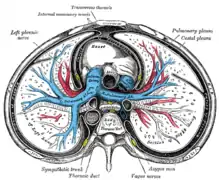
 Transverse section of thorax, showing relations of pulmonary artery.
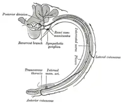
 Diagram of the course and branches of a typical intercostal nerve.
References
This article incorporates text in the public domain from page 403 of the 20th edition of Gray's Anatomy (1918)
External links
- Anatomy photo:18:05-0104 at the SUNY Downstate Medical Center - "Thoracic Wall: Removal of Intercostal Muscles"
- thoraxmuscles at The Anatomy Lesson by Wesley Norman (Georgetown University)