Tricholoma vaccinum
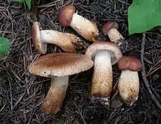
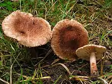
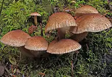
Tricholoma vaccinum, commonly known as the russet scaly tricholoma, the scaly knight, or the fuzztop, is a fungus of the agaric genus Tricholoma. It produces medium-sized fruit bodies (mushrooms) that have a distinctive hairy reddish-brown cap with a shaggy margin when young. The cap, which can reach a diameter of up to 6.5 cm (2.6 in) wide, breaks up into flattened scales in maturity. It has cream-buff to pinkish gills with brown spots. Its fibrous, hollow stipe is white above and reddish brown below, and measures 4 to 7.5 cm (1.6 to 3.0 in) long. Although young fruit bodies have a partial veil, it does not leave a ring on the stipe.
| Tricholoma vaccinum | |
|---|---|
 | |
| Scientific classification | |
| Kingdom: | |
| Division: | |
| Class: | |
| Order: | |
| Family: | |
| Genus: | |
| Species: | T. vaccinum |
| Binomial name | |
| Tricholoma vaccinum | |
| Synonyms[1] | |
| Tricholoma vaccinum | |
|---|---|
float | |
| gills on hymenium | |
| cap is convex or flat | |
| hymenium is adnate or sinuate | |
| stipe is bare | |
| spore print is white | |
| ecology is mycorrhizal | |
| edibility: edible but not recommended | |
Widely distributed in the Northern Hemisphere, Tricholoma vaccinum is found in northern Asia, Europe and North America. The fungus grows in a mycorrhizal association with spruce or pine trees, and its mushrooms are found on the ground growing in groups or clusters in late summer and autumn. Although some consider the mushroom edible, it is of poor quality and not recommended for consumption. The ectomycorrhizae of T. vaccinum has been the subject of considerable research.
Taxonomy and naming
The species was first described in 1774 by German mycologist Jacob Christian Schäffer as Agaricus vaccinus.[6] According to MycoBank, synonyms include August Batsch's 1783 Agaricus rufolivescens, Jean-Baptiste Lamarck's 1783 Amanita punctata var. punctata, and Lucien Quélet's 1886 Gyrophila vaccina.[1] Marcel Bon described the variety T. vaccinum var. fulvosquamosum in 1970, which has squamules (minute scales) arranged in a concentric fashion on the cap;[7] Manfred Enderle published this taxon as a form in 2004.[8]
According to the infrageneric classification of Tricholoma proposed by Rolf Singer in 1986,[9] Tricholoma vaccinum is placed in the section Imbricata, subgenus Tricholoma in the genus Tricholoma. Imbricata includes species with a dry cap cuticle, with a texture that ranges from roughened or squamulose (resembling suede) to almost smooth.[10] The specific epithet derives from the Latin word vaccinus and means "cow-colored". The mushroom's common names include the "russet scaly tricholoma",[11] "fuzztop",[12] and "scaly knight".[13]
Description

The cap of T. vaccinum is initially broadly conical, then convex and finally flattened; its diameter is usually between 2.5 and 6.5 cm (1.0 and 2.6 in). The cap margin is initially curved inwards, and shaggy from hanging remnants of the partial veil. The partial veil is cotton-like, and does not leave a ring on the stipe. The fibrous to scaly cap surface ranges in color from reddish-cinnamon to brownish-orange to tan. The gills have an adnate to sinuate attachment to the stipe, and are crowded closely together. There are between three and nine tiers of lamellulae—short gills that do not extend completely from the cap edge to the stipe. The gills are dingy white, and frequently stain reddish brown. The stipe is 4 to 7.5 cm (1.6 to 3.0 in) long and 1 to 2.2 cm (0.4 to 0.9 in) thick, and becomes hollow in age. It is roughly equal in width throughout its length and ranges in color from whitish to the same color as the cap, but lighter, and sometimes with reddish-brown stains; it is lighter in color near the apex. Like the cap, the stipe surface is fibrous to scaly.[14] The odor of the fruit bodies is unpleasant.[15]
The mushrooms produce a white spore print, and the spores are broadly elliptical, smooth, hyaline (translucent), inamyloid, measuring 6–7.5 by 4–5 μm.[11] The basidia (spore-bearing cells) are four-spored, without clamps, and measure 17–32 by 6.0–7.5 μm. The hymenium lacks cystidia. The arrangement of the hyphae in the cap cuticle ranges from a cutis (with hyphae that run parallel to the cap surface) to a trichoderm (hyphae perpendicular to the cap surface); these hyphae are roughly cylindrical, and measure 3.5–8.0 μm wide, with roughly cylindrical to club-shaped ends that are 6.0–11.0 μm wide. There are no clamp connections in the hyphae of T. vaccinum.[14]
Although the fruit bodies are considered edible,[16] they are of low quality, and generally not recommended for consumption due to their resemblance to and potential for confusion with toxic brown Tricholomas.[17] Orson K. Miller, Jr. considers them "bitter and not edible".[15] Roger Phillips says they may be poisonous.[5] The fruit bodies can be used to create yellow dyes to color wool or other fibers.[18]
Similar species

With its reddish-brown wooly cap, white gills, and hollow stipe, Tricholoma vaccinum is a fairly distinct mushroom and is unlikely to be confused with other Tricholoma.[19] Tricholoma imbricatum somewhat resembles T. vaccinum, but has duller brown colors, is less robust in stature, and has a solid (not hollow) stalk.[12] Another lookalike, T. inodermeum, has a less woolly cap texture and flesh that turns bright pinkish red when injured. It associates solely with pine species and prefers calcareous soil.[14] Other brownish Tricholoma species include T. fracticum, T. dryophilum, and T. muricatum.[20] The scaly and fibrous cap surface of T. vaccinum might be confused with Inocybe, but species in this genus can be distinguished by their brown spore prints.[21]
Habitat and distribution

Tricholoma vaccinum is a mycorrhizal species, and grows in association with coniferous trees, especially pine and spruce.[17] It forms ectomycorrhizae that have been called "Medium-Distance fringe exploration type", indicative of the ectomycorrhiza's ecological role in space occupation in the soil, their possible reach regarding nutrient acquisition and their demand of carbohydrates that have to be invested by the trees for their fungal partners.[22] Fruit bodies usually appear in groups or clusters on the ground, sometimes with moss. The fungus fruits in late summer and autumn.[17] It is found in northern Asia,[13] Europe, and, in North America, is widely distributed in the United States and Canada,[12] and has also been recorded in Mexico.[23] It is one of the most common species of Tricholoma in Central Europe,[24] and is often found in large groups in spruce forests.[19] It is rare in the United Kingdom, and most records have been from Scotland.[10] The fungus may be extinct in the Netherlands.[13]
The ectomycorrhizae of T. vaccinum has been the subject of considerable research. Ectomycorrhizae of Tricholoma species can vary considerably among species in the genus, and differences in the structure of rhizomorphs (a cordlike fusions of hyphae resembling a root) have been used to key out species.[25] Mycorrhizae formed with Norway spruce (Picea abies) are conspicuously hairy with numerous hyphae. The hyphae are partly densely interconnected to rhizomorphs that have a pigment in their outer membrane. The emanating hyphae mostly lack "contact septae" (fully developed simple septae) and contact clamps, and the rhizomorph hyphae vary markedly in diameter. The Hartig net (a network of hyphae that extend into the root) formed by T. vaccinum grows more deeply towards the epidermis, is composed of more rows of hyphae and has more tannin cells in close proximity to the epidermis, and consequently, fewer cortical cells in this position. It is here that the rhizomorphs make the closest contact with the rootlets.[26] The mantle is prosenchymatous, meaning that the constituent hyphae are loosely organized with spaces between them.[25] A combination of techniques including freeze fracturing and scanning electron microscopy has been used to probe the microstructure of the ectomycorrhizae, including inner mantle thickness and the nature of the interface between the Hartig net and host cells.[27] Several fungal genes specifically expressed during ectomycorrhizal interaction between T. vaccinum and Picea abies have been identified, including some involved in a plant pathogen response, nutrient exchange and growth in the plant, signal transduction, and stress response.[28] The first characterized fungal aldehyde dehydrogenase enzyme, ALD1, helps circumvent ethanol stress—a critical function in mycorrhizal habitats.[29]
See also
- List of North American Tricholoma
- List of Tricholoma species
References
- "Tricholoma vaccinum (Schaeff.) P. Kumm. 1871". MycoBank. International Mycological Association. Retrieved 2011-12-26.
- Batsch AJGK (1783). "Elenchus fungorum" (in Latin and German). Halle-Magdeburg, Germany: Joannem J. Gebaver: 85. Cite journal requires
|journal=(help) - Persoon CH (1798). Icones et Descriptiones Fungorum Minus Cognitorum (in Latin). 1. Leipzig, Germany: Bibliopolii Breitkopf-Haerteliani impensis. p. 6.
- Quélet L. (1886). Enchiridion Fungorum in Europa media et praesertim in Gallia Vigentium (in Latin). France: Octavii Doin. p. 12.
- Phillips, Roger (2010). Mushrooms and Other Fungi of North America. Buffalo, NY: Firefly Books. p. 49. ISBN 978-1-55407-651-2.
- Schäffer JC (1774). Fungorum qui in Bavaria et Palatinatu circa Ratisbonam nascuntur Icones (in Latin). 4. Erlangen, Germany: Apud J.J. Palmium. p. 13.
- Bon M. (1969). "Révision des Tricholomes" (in French). 85: 475–92. Cite journal requires
|journal=(help) - Enderle M. (2004). Die Pilzflora des Ulmer Raumes (in German). Ulm, Germany: Verein für Naturwiss. u. Mathematik. pp. 1–521 (see p. 279). ISBN 978-3-88294-336-8.
- Singer R. (1986). The Agaricales in Modern Taxonomy (4th ed.). Königstein im Taunus, Germany: Koeltz Scientific Books. ISBN 978-3-87429-254-2.
- Kibby G. (2010). "The genus Tricholoma in Britain". Field Mycology. 11 (4): 113–40. doi:10.1016/j.fldmyc.2010.10.004.
- Roody WC (2003). Mushrooms of West Virginia and the Central Appalachians. Lexington, Kentucky: University Press of Kentucky. p. 264. ISBN 978-0-8131-9039-6.
- McKnight VB, McKnight KH (1987). A Field Guide to Mushrooms: North America. Peterson Field Guides. Boston, Massachusetts: Houghton Mifflin. p. 191. ISBN 978-0-395-91090-0.
- Roberts P, Evans S (2011). The Book of Fungi. Chicago, Illinois: University of Chicago Press. p. 316. ISBN 978-0-226-72117-0.
- Bas C, Noordeloos ME, Kuyper TW, Vellinga EC (1999). Flora Agaricina Neerlandica. 4. Rotterdam, Netherlands: A.A. Balkema Publishers. p. 121. ISBN 978-90-5410-493-3.
- Miller HR, Miller OK (2006). North American Mushrooms: A Field Guide to Edible and Inedible Fungi. Guilford, Connecticut: Falcon Guide. p. 132. ISBN 978-0-7627-3109-1.
- Dickinson C, Lucas J (1982). VNR Color Dictionary of Mushrooms. New York, New York: Van Nostrand Reinhold. p. 157. ISBN 978-0-442-21998-7.
- Ammirati J, Trudell S (2009). Mushrooms of the Pacific Northwest. Timber Press Field Guides. Portland, Oregon: Timber Press. p. 107. ISBN 978-0-88192-935-5.
- Bessette A, Bessette AR (2001). The Rainbow Beneath my Feet: A Mushroom Dyer's Field Guide. Syracuse, New York: Syracuse University Press. p. 163. ISBN 978-0-8156-0680-2.
- Noordeloos ME, van Zanen G (2005). "De Ruige ridderzwam in de Flevopolder" [Tricholoma vaccinum in the Flevopolders] (PDF). Coolia (in Dutch). 48 (1): 18–9.
- Davis RM, Sommer R, Menge JA (2012). Field Guide to Mushrooms of Western North America. Berkeley, California: University of California Press. p. 168. ISBN 978-0-520-95360-4.
- Lincoff G. (1981). The Audubon Society Field Guide to North American Mushrooms. New York, New York: Knopf. pp. 805–6. ISBN 978-0-394-51992-0.
- Agerer R. (2007). "Diversitat der Ektomykorrhizen im unter- und oberirdischen Vergleich: die Explorationstypen" [Diversity of ectomycorrhizae as seen from below and above ground: the exploration types]. Zeitschrift für Mykologie (in German and English). 73 (1): 61–88. ISSN 0170-110X.
- De Avila B, Welden AL, Guzmán G (1980). "Notes on the ethnopharmacology of Hueyapan, Morelos, Mexico". Journal of Ethnopharmacology. 2 (4): 311–21. doi:10.1016/S0378-8741(80)81013-0. PMID 7421279.
- Pilát, Albert (1961). Mushrooms and other Fungi. London, UK: Peter Nevill. p. 71.
- Brunner I, Amiet R, Zollinger M, Egli S (1992). "Ectomycorrhizal synthesis with Picea abies and three fungal species: a case study on the use of an in vitro technique to identify naturally occurring ectomycorrhizae". Mycorrhiza. 2 (2): 89–96. doi:10.1007/BF00203255. S2CID 24671418.
- Agerer R. (1987). "Studies on ectomycorrhizae .9. Mycorrhizae formed by Tricholoma sulfureum and Tricholoma vaccinum on spruce". Mycotaxon. 28 (2): 327–60.
- Scheidegger C, Brunner I (1993). "Freeze-fracturing for low-temperature scanning electron-microscopy of Hartig net in synthesized Picea abies, Hebeloma crustuliniforme and Tricholoma vaccinum ectomycorrhizas". New Phytologist. 123 (1): 123–32. doi:10.1111/j.1469-8137.1993.tb04538.x. JSTOR 2557778.
- Krause K.; Kothe (2006). "Use of RNA fingerprinting to identify fungal genes specifically expressed during ectomycorrhizal interaction". Journal of Basic Microbiology. 46 (5): 387–99. doi:10.1002/jobm.200610153. PMID 17009294.
- Asiimwe T, Krause K, Schlunk I, Kothe E (2012). "Modulation of ethanol stress tolerance by aldehyde dehydrogenase in the mycorrhizal fungus Tricholoma vaccinum". Mycorrhiza. 22 (6): 471–84. doi:10.1007/s00572-011-0424-9. PMID 22159964. S2CID 14809764.
External links
| Wikimedia Commons has media related to Tricholoma vaccinum. |