Valonia ventricosa
Valonia ventricosa, also known as bubble algae or sailor's eyeballs,[2] is a species of alga found in oceans throughout the world in tropical and subtropical regions. It is one of the largest unicellular organisms, if not the largest.[2][3]

| Valonia ventricosa | |
|---|---|
 | |
| Scientific classification | |
| Phylum: | Chlorophyta |
| Class: | Ulvophyceae |
| Order: | Cladophorales |
| Family: | Valoniaceae |
| Genus: | Valonia |
| Species: | V. ventricosa |
| Binomial name | |
| Valonia ventricosa | |
| Synonyms | |
|
Ventricaria ventricosa | |
Characteristics
Valonia ventricosa has a coenocytic structure with multiple nuclei and chloroplasts. This organism possesses a large central vacuole which is multilobular in structure (lobules radiating from a central spheroid region).
The entire cell contains several cytoplasmic domains with each domain having a nucleus and a few chloroplasts. Cytoplasmic domains are interconnected by cytoplasmic "bridges" that are supported by microtubules. The peripheral cytoplasm (whose membrane is overlaid by the cell wall), is only about 40 nm thick.
Valonia ventricosa typically grow individually, but in rare cases they can grow in groups.
Environment
They appear in tidal zones of tropical and subtropical areas, like the Caribbean, north through Florida, south to Brazil, and in the Indo-Pacific.[2] Overall, they inhabit every ocean throughout the world,[4] often living in coral rubble.[5] The greatest observed depth for viability is approximately 80 metres (260 ft).
Physiology and reproduction
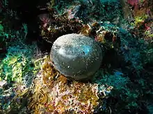
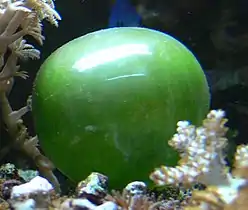
The single-cell organism has forms ranging from spherical to ovoid, and the color varies from grass green to dark green, although in water they may appear to be silver, teal, or even blackish.[2] This is determined by the quantity of chloroplasts of the specimen.[5] The surface of the cell shines like glass when clean due to being extremely smooth with no texture. The thallus consists of a thin-walled, tough, multinucleate cell with a diameter that ranges typically from 1 to 4 centimetres (0.4 to 1.6 in) although it may achieve a diameter of up to 5.1 centimetres (2.0 in) in rarer cases. The "bubble" alga is attached by rhizoids to the substrate fibers.[2]
Reproduction occurs by segregative cell division, where the multinucleate parent cell makes child cells, and individual rhizoids form new bubbles, which become separate from the parent cell.
Studies
Valonia ventricosa has been studied particularly because the cells are so unusually large that they provide a convenient subject for studying the transfer of water and water-soluble molecules across biological membranes. It was concluded that the properties of permeability in both osmosis and diffusion were identical, and that urea and formaldehyde molecules did not require any kind of postulated water-filled pores in the membrane to move through it.[2][6][7] In studying the cellulose lattice, and its orientation in biological structures, Valonia ventricosa has undergone extensive X-ray analytical procedures.[8] It has also been studied for its electrical properties, due to its unusually high electrical potential relative to the seawater that surrounds it.[6]
See also
References
- "Valonia ventricosa J. Agardh". ITIS. Retrieved 27 August 2010.
- Bauer, Becky (October 2008). "Gazing Balls in the Sea". All at Sea. Retrieved 26 September 2013.
- Tunnell, John Wesley; Chávez, Ernesto A.; Withers, Kim (2007). Coral reefs of the southern Gulf of Mexico. Texas A&M University Press. p. 91. ISBN 978-1-58544-617-9.
- "Valonia ventricosa J.Agardh", Algaebase, retrieved 4 September 2015
- Lee, Robert Edward (2008). "Siphonoclades". Phycology. Cambridge University Press. p. 189. ISBN 978-0-521-68277-0. Retrieved 27 August 2010.
- Thellier, M. (1977). Échanges ioniques transmembranaires chez les végétaux. Publication Univ Rouen Havre. p. 341. ISBN 978-2-222-02021-9.
- Gutknecht, John (1967). "Membranes of Valonia ventricosa: Apparent Absence of Water-Filled Pores". Science. Science Magazine. 158 (3802): 787–788. doi:10.1126/science.158.3802.787.
- Astbury, W. T.; Marwick, T. C.; Bernal, J. D. (1932). "X-Ray Analysis of the Structure of the Wall of Valonia ventricosa.--I". Proceedings of the Royal Society of London. Series B. 109 (764): 443. doi:10.1098/rspb.1932.0005. JSTOR 81568.
External links
- Wardrop, A. B.; Jutte, S. M. (1968). "The enzymatic degradation of cellulose from Valonia ventricosa". Wood Science and Technology. 2 (2): 105. doi:10.1007/BF00394959.
- Revol, J (1982). "On the cross-sectional shape of cellulose crystallites in Valonia ventricosa". Carbohydrate Polymers. 2 (2): 123–134. doi:10.1016/0144-8617(82)90058-3.
- Friday Fellow: Sailor's Eyeball at Earthling Nature